Dental Cavities
The tooth and nothing but the tooth (well almost)
Dental cavities in the enamel and dentin areas of a tooth can be a significant problem. However, it can be prevented to a great degree if you understand its causes and practice a few basic techniques.
Dental Cavities
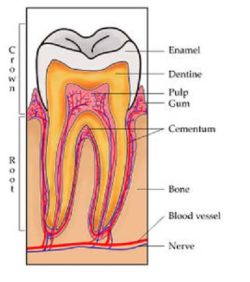
Tooth Composition: Enamel and Dentin
|
The majority of a tooth's surface that is above the gum line is made up of enamel. Tooth enamel is the hardest tissue found in the human body. Enamel is composed of more than 95% mineral content which is rich in calcium. |
 |
Dental Cavities Table of Contents
- Cavities - The Breakdown
- Classifying Cavities
- Signs and Symptoms of Cavities
- Diagnosing Cavities
- Causes of Cavities
- Other Risk Factors
- Treating Cavities
- Preventive Techniques (Includes Video)
- Facts and Recommendations for Managing Cavities
Dental Cavities - The Breakdown
|
Dental Cavities are located on a tooth where a hole is formed because so much of the tooth's mineral content has dissolved away. This hole is an infectious disease which can damage the structure of a tooth. It can lead to pain, tooth loss, further infection, and in severe cases, death. |
 |
Two terms are used interchangeable when referring to dental cavities. One is dental caries, the other is tooth decay. Dental caries is the term most often used by dental providers. It is also a word derived from the Latin word for "rot," - an appropriate description for the tooth decaying process. And while there exist several ways to classify dental cavities, the risk factors and development process remain the same.
Dental cavities are caused by certain bacterial plaque acid. Bacteria causes the tooth’s surface to demineralize (lose its mineral content) and causes the most damage in the presence of sugar. The bacteria which contribute to tooth decay are living organisms, just like us. They reside inside our mouths and consume the food we eat, and create waste products in the form of acids. Sugars (glucose, sucrose, fructose, lactose, or cooked starches) are their food source. The waste products they create is the acids, such as lactic acid, which cause demineralization of tooth enamel. This process begins within minutes of our ingesting foods which contain sugars.
Note: Everyone's mouth contains bacteria. In fact, a single human mouth can contain more microorganisms than there are people on planet Earth. While we cannot sterilize our mouths, we can minimize the potential for getting tooth decay by prevent the bacteria that is present from forming bacterial colonies. These bacterial colonies are better known as dental plaque.
With time, saliva can penetrate through the dental plaque and begin to neutralize the acids that were created in response to the consumption of sugar. Unfortunately, this can take as long as two or more hours.
The amount of tooth demineralization that takes place after an exposure to sugar is partly related to the age of the dental plaque. Given the same exposed to sugar, dental plaque that is only a few hours old will not produce as much tooth demineralization as dental plaque that is several days old.
When conditions at a tooth's surface are non-acidic, mineral build up (remineralization) can take place. During this process, minerals in the mouth can be re-incorporated into a tooth, thus reversing, or at least minimizing, the damage that was done to the tooth during the tooth decay process (demineralization). This repair process will either continue until the repair has been completed, or be interrupted by the next attack of bacterial acids.
This tug of war between the mineral loss and mineral buildup processes, which occurs several times a day, is a reason why dental cavities can take many months to form. In some cases, a tooth's mineral build up can balance out the damage done during a tooth’s mineral loss stage. However, in cases where there is heavy dental plaque accumulation or the person has a high sugar intake, the balance will tip in favor of tooth decay formation.
Saliva plays a very important role in process of fighting dental cavities. It contains buffering agents that can neutralize the bacterial acids found in dental plaque and contains minerals for the remineralization process to occur. Saliva also contains antibacterial agents that can inhibit the growth of oral bacteria. A reduction of saliva reduces the benefits it provides in fighting tooth decay. This condition is known as xerostomia - a state of diminished salivary flow. It is often caused by aging whereby the salivary glands become less effective or a change in the saliva composition, placing us more at risk for tooth decay. Xerostomia can also be associated as a side effect for a person taking medication. Antihistamines (allergy and cold medications), antidepressants, blood pressure agents, and anti-anxiety medications are some known medicines which cause mouth dryness.
Classifying Dental Cavities
Dental cavities are classified as follows:
- Dental Cavities Classified by Location
- Dental Cavities Classified by Other General Locations
- Dental Cavities Classified by Etiology
- Dental Cavities Classified by Rate of Progression
- Dental Cavities Classified by Affected Hard Tissue
When used to characterize a particular case of tooth decay, using classifications more accurately represents the condition to others and may also indicate the severity of tooth destruction.
I. Dental Cavities by Location
Generally, there are two types of cavities when separated by location: Smooth Surface Caries and Pits & Fissures Caries. The location, development, and progression of smooth-surface caries differ from those of pit and fissure caries.
Smooth Surface Dental Cavities are found on smooth surfaces of the teeth. There are three types of smooth surface caries: Proximal or Interproximal, Root Caries and All Other Smooth Surfaces.
Proximal Caries
| Proximal caries form on the smooth surfaces between adjacent teeth. Proximal caries are the most difficult to detect. It cannot be detected visually or manually with a dental explorer, so an x-ray is needed for early discovery. | 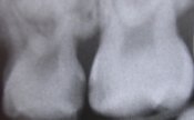 Wikipedia photograph taken of radiograph showing interproximal decay between first and second left maxillary first molars (i and j) |
Root Caries
Root caries form on the root surfaces of teeth. Root caries are the third most common type of caries and usually occur when the root surfaces have been exposed to bacterial plaque.
The root portion of a tooth is neither composed of nor covered over by dental enamel. It is primarily made up of a softer mineralized tooth tissue called dentin. Dentin contains less mineral content than enamel and as such, is more easily damaged by the demineralization process. If gum recession occurs, the tooth's root is exposed and because the location of its surface, plaque is continually present and decay forms more readily.
Gum recession happens for a number of reasons. Someone with gum disease - most often associated with a persistent accumulation of dental plaque, who brushes improperly, (very hard) or brushes with a toothbrush that is very bristle, or constantly grinds and clenches the teeth, is susceptible to getting gum recession. As we get older, a culmination of these factors and others, increases the amount of gum recession that is present in our mouths.
Although root caries is more susceptible to tooth decay than proximal caries because of the softer material composition of dentin, it is easier to stop the progression of root caries than enamel caries. This is because the roots of teeth have a greater reuptake of fluoride than enamel.
Root caries are most likely to be found in the following order: on facial surfaces, then interproximal surfaces, then lingual surfaces.
Lower (mandibular) molars are the most common location to find root caries, followed by:
-
- lower (mandibular) premolars,
- upper (maxillary) front (anteriors) teeth,
- upper (maxillary) back (posterior) teeth, then
- lower (mandibular) front (anterior) teeth.
All Other Smooth Surface Caries
Smooth surface caries are dental cavities which occur on any other smooth tooth surface other than the interproximal area. Because these caries occur in all smooth surface areas of enamel except for interproximal areas, they are easily detected.
Fissures and Pits Caries
| Fissures are grooves located on the chewing surfaces of back teeth and lingual surfaces of upper front teeth. Pits are small, pinpoint depressions that are found at the ends or cross-sections of grooves. Buccal pits are found on the facial surface of molars. For all types of pits and fissures, the deep infolding of enamel makes oral hygiene along these surfaces difficult, allowing dental cavities to be common in these areas. | 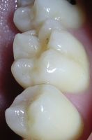 |
II. Other General Caries Descriptions
Besides smooth surface caries and fissure and pits caries, carious lesions can be described further by their location on a particular surface of a tooth: Facial, Lingual, Cervical, Occlusal, Incisal, Mesial and Distal.
- Facial Caries - caries on a tooth's surface that are nearest to the cheeks or lips. Facial caries can be subdivided into two areas:
- Lingual Caries -caries on surfaces facing the tongue. Lingual caries can also be described as palatal when found on the lingual surfaces of upper teeth because they are located beside the hard palate.
- Cervical Caries - found near a tooth's cervix—the location where the crown of a tooth and its roots meet.
- Occlusal Caries - found on the chewing surfaces of back teeth.
- Incisal Caries -found on the chewing surfaces of anterior teeth.
- Mesial Caries - is a location on a tooth closer to the median line of the face, which is located on a vertical axis between the eyes, down the nose, and between the contact of the central incisors.
- Distal Caries - the locations on a tooth further away from the median line.
-
- buccal (found on the surfaces of posterior teeth nearest the cheeks)
- labial (found on the surfaces of anterior teeth nearest the lips).
III. Dental Cavities by Etiology
In some instances, caries are described in ways that might indicate the cause. "Baby bottle caries", "early childhood caries", or "baby bottle tooth decay" is a pattern of decay found in young children with their baby teeth. The teeth most likely affected are the upper front teeth, but all teeth can be affected. The name for this type of caries comes from the fact that the decay usually is a result of allowing children to fall asleep with sweetened liquids in their bottles or feeding children sweetened liquids multiple times during the day.
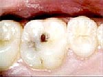
Example of Rampant Caries
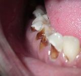 |
Another pattern of decay is "rampant caries", which signifies advanced or severe decay on multiple surfaces of many teeth. |
IV. Dental Caries by Rate of Progression
Descriptions are applied to caries to indicate the progression rate and previous history.
- "Acute" signifies a quickly developing condition
- "Chronic" describes a condition which has taken an extended time to develop.
- Recurrent caries is caries that appear at a location with a previous history of caries. This is frequently found on the margins of fillings and other dental restorations.
- Incipient caries describes decay at a location that has not experienced previous decay.
- Arrested caries describes a lesion on a tooth which was previously demineralized but was remineralized before causing a cavitation.
V. Dental Cavities Classified by Affected Hard Tissue
Depending on which hard tissues are affected, it is possible to describe caries as involving enamel, dentin, or cementum - a bony layer of connective tissue that covers the dentin part of a tooth.
Early in its development, dental cavities may affect only enamel. Once the extent of decay reaches the deeper layer of dentin, "dentinal caries" is used to describe the caries.
Since cementum is the hard tissue that covers the roots of teeth, it is not often affected by decay unless the roots of teeth are exposed to the mouth. Although the term "cementum caries" may be used to describe the decay on roots of teeth, very rarely does caries affect the cementum alone. Roots have a very thin layer of cementum over a large layer of dentin, and thus most caries affecting cementum also affects dentin.
Signs and Symptoms of Dental Cavities
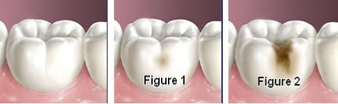
Until dental cavities progresses, a person may not be aware of it. The earliest sign of a new carious lesion is the appearance of a chalky white spot on the surface of the tooth which indicates demineralization of the enamel (Figure 1). As the lesion continues to demineralize, it will eventually turn into a dental cavity (Figure 2). The process before this point (Figure 2) is reversible, but once a cavity forms, the lost tooth structure cannot be regenerated.
As the enamel and dentin are destroyed further, dental cavities become more noticeable. The affected areas of the tooth change color and become soft to the touch. Once the decay passes through enamel, the dentinal tubules, which contain passages to the nerve of the tooth, become exposed and cause the tooth to hurt. Dental cavities can also cause bad breath and foul tastes. In highly progressed cases, infection can spread from the tooth to the surrounding soft tissues and become life-threatening.
Diagnosing Dental Cavities
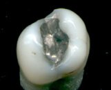
| Primary diagnosis of dental cavities involves inspecting all visible tooth surfaces using a good light source, dental mirror, explorer and dental radiographs (x-rays). (See a basic examination procedure on video below). The use of x-rays can reveal dental caries in a patient's mouth before it is otherwise visible, particularly in the case of caries on interproximal (between the teeth) surfaces. Large dental caries are often apparent to the naked eye, but smaller lesions can be difficult to identify. Below is a cavity that has been diagnosed very early, and is therefore, being treated without the use of needles or the dental drill. Talk about making certain to have regular dental checkups! |  |
When a dental radiograph is taken, the hard mineralized tooth tissues block out some of the x-rays attempting to pass through the tooth on their way to reaching the x-ray film. So, the parts of the dental film that lie protected behind a tooth's enamel and dentin portions will have less exposure and look lighter in color on the x-ray film. Since the areas of tooth decay are a demineralization of the tooth's hard tissues, or a hole in the tooth, if the decay process has progressed far enough, those locations on the tooth where decay has formed will appear as darkened areas on a radiograph. This is because the decayed portion of the tooth is less "hard," therefore, the x-rays can penetrate that portion of the tooth more easily and expose the dental film to a greater degree. Therefore, when a dentist evaluates a dental x-ray for evidence of tooth decay, they are examining the image of the teeth on the film for hints of changes in the density of the tooth's enamel and dentin.
In the past, small dental cavities were found by searching for soft areas of tooth structure with a dental explorer. Some dental researchers have since cautioned against the use of dental explorers to find dental cavities. This is because in cases where a small area of tooth has begun demineralizing, but has not yet cavitated, the pressure from the dental explorer could cause a cavitation. Since the carious process is reversible before a cavitation is present, it may be possible to stop the caries with fluoride to remineralize the tooth surface. If cavitation is present, a restoration is the only option to replace the lost tooth structure.
An alternative technique that is proposed for determining the presence of cavities is by using air. Air that is blown across a tooth's surface removes moisture and changes the optical properties of the unmineralized enamel. This process produces a white 'halo' effect that is detectable to the naked eye. The use of fiberoptic transillumination, lasers and disclosing dyes have also been recommended for use as an adjunct when diagnosing smaller carious lesions in pits and fissures of teeth.
Depending on the extent of tooth destruction, various treatments can be used to restore teeth to proper form, function, and esthetics. Unfortunately, there is no known method to regenerate large amounts of tooth structure.
Causes of Dental Cavities
There are four main criteria required for dental cavities formation: a tooth surface, acid producing bacteria, sugar and time. Each item is explained below.
-
Tooth Surface
Because the process of dental cavities does not have an inevitable outcome, individuals will be susceptible to different degrees of carious lesions. This is dependent upon such things as the shape of their teeth, their oral hygiene habits, and the buffering capacity of their saliva. The anatomy of teeth may affect the likelihood of caries formation. In cases where the deep grooves of teeth are more numerous and exaggerated, pit and fissure caries are more likely to develop.
Bacteria
The mouth contains a wide variety of bacteria, but only a few specific species of bacteria are believed to cause dental caries. This bacteria forms as plaque in the mouth, yet some areas of the mouth collect plaque more commonly than other areas. For example, the grooves on the biting surfaces of molar and premolar teeth, and the point of contact between teeth provide for bacterial retention. Plaque may also collect along the gingiva. In addition, the edges of fillings or crowns can provide protection for bacteria, as can intraoral appliances such as orthodontic braces or removable partial dentures.
Sugar
In the mouth, bacteria changes sugars into acids. If left in contact with the tooth, these acids cause demineralization. The process is dynamic, however, as remineralization can also occur if the acid is neutralized and suitable minerals are available in the mouth from saliva and preventative aids such as fluoride toothpaste and mouthwash. Dental caries may be arrested at this stage.
Time
If sufficient acid is produced over a period of time without remineralization, caries will progress. It may result in so much mineral content being lost that the soft organic material left behind will disintegrate, forming a cavity or hole. Therefore, the frequency with which teeth are exposed to acidic environments increases the likelihood of caries development.
The acid which formed from bacteria eating on meals or snacks which contain sugar causes the pH level in the mouth to decrease. As time progresses, the pH returns to normal due to the buffering capacity of saliva and the dissolved mineral content from tooth surfaces. But during every exposure to the acidic environment, portions of the inorganic mineral content at the surface of teeth dissolve. They can remain dissolved for about 2 hours which is enough time for the tooth surface to be vulnerable to an acidic environment which is also enough time for dental caries to begin to develop. Therefore, the more frequently you consume sugar, the greater your chances of developing a dental cavity.
To sum it up, when sugars are eaten continuously throughout the day, the tooth becomes more and more vulnerable to caries for a longer period of time. This is because the pH is never given an opportunity to returns to normal levels and allow the tooth surfaces to remineralize, or regain its lost mineral content. Proximal caries progress quicker than smooth surface caries. It can take proximal caries an average of 4 years to pass through enamel in permanent teeth. Because the cementum enveloping the root surface is not nearly as durable as the enamel encasing the crown, root caries tends to progress much more rapidly than decay on other surfaces. The progression and loss of mineralization on the root surface is 2.5 times faster than caries in enamel.
Other Risk Factors
In addition to the four main requirements for caries formation, a reduction in a person's saliva can also result in an increased caries rate. Lack of saliva, which acts as a buffering agent, results in an inability to counterbalance the acidic environment created by certain foods. As a result, medical conditions that reduce the amount of saliva produced by salivary glands, particularly the parotid gland, are likely to cause widespread tooth decay.
The use of tobacco may also increase the risk for dental cavities formation. Smokeless tobacco frequently contains high sugar content in some brands, possibly increasing the susceptibility to caries. Tobacco use is also a significant risk factor for periodontal disease, which can allow the gingiva to recede. As the gingiva loses attachment to the teeth, the root surface becomes more visible in the mouth. If this occurs, root caries is a concern since the cementum covering the roots of teeth is more easily demineralized by acids in comparison to enamel.
Currently, there is not enough evidence to support a causal relationship between smoking and coronal caries, but there is suggestive evidence of a causal relationship between smoking and root-surface caries.
Treating Dental Caries
 |
Destroyed tooth structure does not fully regenerate, although remineralization of very small carious lesions may occur if dental hygiene is kept at optimal level. For the small lesions, topical fluoride is sometimes used to encourage remineralization. For larger lesions, the progression of dental caries can be stopped by treatment. Therefore, the goal of treatment is to preserve tooth structures and prevent further destruction of the tooth. Generally, early treatment is less painful and less expensive than treatment of extensive decay. |
A dental hand piece is used to remove large portions of decayed material from a tooth. A spoon is a dental instrument used to remove decay carefully and is sometimes employed when the decay in the dentin area of the tooth reaches near the pulp. Once the decay is removed, the missing tooth structure may require a dental filling restoration, root canal and dental crown to restore the tooth to function and aesthetics. Click here to see a painfree filling procedure, provided courtesy of YouTube.
An extraction can also serve as treatment for dental cavities. The removal of the decayed tooth is performed if the tooth is too far destroyed from the decay process for effective restoration. Extractions are sometimes considered if the tooth lacks an opposing tooth or will probably cause further problems in the future, as is often the case with wisdom teeth. Extractions may also be preferred by patients unable or unwilling to undergo the expense or difficulties in restoring the tooth.
Preventive Techniques
| The primary focus of brushing and flossing is to remove and prevent the formation of bacteria forming plaque. As the amount of bacterial plaque increases, the tooth becomes more vulnerable to dental cavities. A toothbrush can be used to remove plaque on most surfaces of the teeth except for areas between teeth. When used correctly, dental floss removes plaque from areas which could otherwise develop proximal caries. See the videos on proper brushing and flossing techniques. |  |
Regular dental examinations and cleanings are also important. Sometimes, complete plaque removal is difficult, and a dentist or dental hygienist may be needed. Along with oral hygiene, radiographs may be taken at dental visits to detect possible dental cavities development in high risk areas of the mouth.
Minimize snacking since it creates a continual supply of nutrition for acid-creating bacteria in the mouth. Brushing after every meal is highly recommended.
Dental Sealants
Dental sealants are a thin plastic-like coating applied to a tooth. Its use is a good means of prevention, as some portions of a tooth can be more at risk for the formation of tooth decay than others. This increased susceptibility is often related to the anatomy of the tooth. In these instances, dental sealants prove to be a valuable aid in helping to prevent dental cavities from beginning.
Some back teeth, especially molars, can be difficult for a person to clean because the grooves in the chewing portion of these teeth (the "pits and fissures") are deep and narrow. Even though the person brushes they are not able to clean dental plaque off effectively because the bristles of their toothbrush are literally too large to scrub the depths of these pits and grooves. Because some plaque perpetually remains, tooth decay can form. A solution for this situation can be the placement of dental sealants.
When a dentist bonds plastic sealant into and over the grooves of a tooth, it results in a surface of the tooth that is flatter and smoother. There are no longer any places on the chewing portion of the tooth that the bristles of a toothbrush can't reach and clean. Since plaque can be removed more easily and effectively, there is a greater chance of preventing the formation of pit and fissure caries, the most common form of dental cavities.
Fluoride Therapy
Fluoride therapy is the most effective dental cavity prevention treatment available today. It has been demonstrated that water fluoridation and fluoride supplements helps in the fight against dental caries, reducing tooth decay between 40 and 70%. Fluoride helps prevent dental decay by binding to crystals in tooth enamel. This makes the enamel more resistant to demineralization and, thus, more resistant to decay. Fluoride has also been show to decrease the rate at which the bacteria in the mouth can produce acids by disrupting their ability to turn sugar into acid. The less sugar that bacteria consumes, the less acidic tooth demineralizing waste they can produce.
Topical fluoride is also recommended to protect the surface of the teeth. This may include a fluoride toothpaste or mouthwash. Many dentists use topical fluoride therapy as part of routine visits. Click here to see if your city's water supply is flouridated. The results are provided by the US Centers for Disease Control and Prevention.
Dental Cavities Summary
As you have seen, dental cavities can cause significant problems, but it can be prevented to a great degree. Below is a summary outlining the facts associated with dental caries and recommendations to practice and develop good oral healthcare. Use these techniques, and if you have any questions, please speak with your dental provider.
Facts and Recommendations for Managing Dental Cavities
- When oral bacteria consume sugars, they start to produce the acids that cause tooth decay within minutes. The less sugar you consume, the fewer number of times you eat sugary foods, and the shorter the period of time that sugars are allowed to remain in your mouth limits the exposure time of your teeth to oral bacteria acids.
Recommendation: Consume less sugar, limit the number of times you eat sugary foods and brush and floss or at least rinse promptly after consuming sugary foods.
- Decay occurs in areas where dental plaque lies on a tooth's surface.
Recommendation: Take the time to be thorough with brushing and flossing. Tooth areas that are not cleaned effectively become susceptible to cavity formation.
- Acid formation and tooth demineralization begin within minutes of the bacteria receiving a sugary meal. It can take up to several hours for saliva to penetrate the layer of dental plaque and neutralize these acids.
Recommendation: Brush and floss promptly after eating to ensure that dental plaque has been cleaned off the surface of your teeth.
- The longer dental plaque has been present on a tooth's surface, the more capable it is of causing tooth damage.
Recommendation: Brush and floss often and effectively. Take the time to be thorough with your brushing and flossing.
- The process of remineralization, which can repair the damage created by the demineralization process, can only occur when a non-acid environment is present. The remineralization process can potentially stabilize the demineralization process.
Recommendation: Promote a non-acidic environment by limiting the amount, frequency, and duration of dietary sugars in your mouth. Minimize the amount of dental plaque which is present by brushing and flossing thoroughly.
- Fluoride helps to provide protection from tooth decay in a variety of ways.
Recommendation: Brush at least three times a day with a toothpaste that contains fluoride. Look for toothpastes that have the American Dental Association's (ADA) "Seal of Approval." This seal indicates that the ADA has evaluated the product and found its fluoride content to be appropriate. Also, drink fluoridated tap water.
- Some portions of a tooth's surface are predisposed to tooth decay formation because their grooves and pits are hard, if not impossible, to clean effectively.
Recommendation: Speak with your dentist about the possibility of dental sealants being placed on those teeth.
- Dry mouth conditions such as xerostomia can increase a person's risk for tooth decay.
Recommendation(s): 1) Because salivary flow decreases when we sleep, be certain to brush and floss thoroughly before going to bed to minimize the presence of dental plaque. 2) If you have chronic dry mouth, drink plenty of water on a frequent basis to stay properly hydrated and to moisten the mouth. 3) Chewing sugarless gum can also be a good way to boost salivary flow. The sugar substitutes found in sugarless gum is not fermented by dental plaque bacteria and therefore will not promote the formation of bacterial acids.
|
|
|
|

Advertising
![]()