Dental Information

On this page, you are provided with some basic dental information. This information should be used with the information provided to you on the many ways for you and your family to get affordable dental work from a health care provider.
Familiarize yourself with this information. It will help you with any treatment planning discussions you may have with your dentist, or if you have a dental emergency.
Dental Information: Tooth Anatomy, Functions and Surface Areas
This area provides dental information on the following:
- The Anatomy of a Tooth - explains the different parts of a tooth.
- Tooth Types and Functions - describes the different types of teeth and the functions they perform.
- Tooth Numbers or Tooth Area in the mouth.
- Tooth Surfaces or Tooth Area in the mouth.
Dental Information: Tooth Anatomy
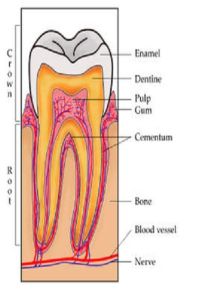
A tooth is a bit like a tree - only part of it is visible, while the roots lie beneath the surface, extending into the gums and into the bones of the mouth. The tooth is made up of many layers: The Crown, Enamel, Cementum, Dentin, Pulp and Root.
The Crown is the top part of the tooth. It is the only part you can normally see. The shape of the crown is determined by its function. For example, front teeth are sharp and chisel-shaped for cutting, while molar teeth have flat surfaces for grinding. The crown can get smaller as we age because the enamel wears down over time. It is usually a gradual process, yet for some people, the process is accelerated due to an incorrect bite or from habits such as grinding the teeth.
The Enamel part of the tooth is the outermost layer of the tooth. Enamel is the hardest tissue in the body as it can withstand the stress of biting, chewing and grinding, but is also brittle and prone to cracking and chipping. Enamel can also be damaged by decay if teeth are not properly cared for.
The layer of tooth underneath the enamel is called the Dentin. Dentin is the bone-like substance that makes up most of the tooth structure. Although white enamel is the outer layer of the tooth, dentin is mostly responsible for the color of the tooth. While coffee, tea, tobacco can cause surface stains in the enamel that can be removed with polishing, there are other types of discoloration which involve the dentin. To change the color of the tooth, the color of the dentin must be changed. And because dentin is a softer material than enamel and when decay progresses its way through the enamel, this is the area where it next attacks. Dentin contains millions of tiny tubes which lead directly to the dental pulp.
Cementum - Just as enamel covers the crown of the tooth, cementum covers the root(s) of the tooth. It is not as hard or as white as enamel. Cementum attaches to tiny fibers that help anchor the tooth to the jawbone.
The Gumline is where the tooth and the gums meet. This is where plaque and tartar can build up if there is no proper brushing and flossing. This will lead to gingivitis, the earliest stage of gum disease.
The Pulp is located in the middle of the tooth and contains blood vessels and nerve fibers that allow you to feel sensations of hot, cold and pain. This is why when tooth decay reaches the pulp, you usually feel pain.
The last item in the tooth is the Root. The root is the part of the tooth that is embedded in bone, as it extends into the upper or lower jawbones. Different types of teeth have different root formations. Incisors and canine teeth have a single root that tapers down from the tooth. Molars may have one, two or three roots depending on their type and location in the mouth. At the end of each root is a small opening that allows blood vessels and nerves to enter the tooth. The root makes up about two-thirds of the tooth and holds the tooth in place.
Understanding the design of a tooth will allow you to:
- more clearly understand what your dental needs are when discussing your treatment plan with an affordable family dental care provider.
- understand what is happening when a dentist emergency occurs. You are in a better position to temporize the problem until you are able to seek affordable dental work from an affordable dentist.
- teach your family how to achieve better oral health. Then, the options of finding affordable family dental care will become greater.
Understanding the design of a tooth places you in a better position to understand your dental needs. If the dental work that you will need is preventative care, there will be no need to have procedures performed by a dental dentist specialist. This places you in a better position to save money on dental work for you and your family.
So, familiarize yourself with dental information provided on the anatomy of the tooth and continue on the road to affordable family dental care by going to the next topic: Tooth Types and their Functions.
Dental Information: Tooth Types and their Functions
There are 4 basic types of teeth. Each type has a specific job or function. They are categorized as Incisors, Canines, Premolars and Molars. Dental information on each is described below.
Incisors - the sharp, chisel-shaped front teeth (four upper, four lower) used for cutting food.
Canines - sometimes called cuspids, these teeth are shaped like points (cusps) and are used for tearing food.
Premolars - these teeth have two pointed cusps on their biting surface and are sometimes referred to as bicuspids. The premolars are for crushing and tearing.
Molars - used for grinding, these teeth have several cusps on the biting surface.
Dental Information: Determining a Tooth Number or Area
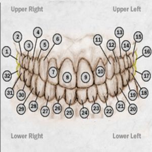
As adults, we have (or should have) a total of 32 teeth. All dentists refer to tooth numbers or areas when describing a situation and procedure with a particular tooth.
The teeth are numbered beginning from the back of the right side of the mouth on the top. For instance, the last tooth all the way in the back on the right-side of the mouth is referred to as tooth number 1. If you proceed forward towards the front and the left side of the mouth, you should count a total of 16 teeth total. If there are missing teeth in your mouth, include the spaces in your count, as this is also considered a tooth area.
After counting 16 teeth or areas on the top of the mouth, you should be on the left side of your mouth. Simply, lower your finger down to the left side bottom and continue to count, continuing at number 17. Proceed across the lower half of the mouth over to the right side until you are able to determine 32 teeth or areas of the mouth.
This is how you determine the tooth area of the mouth. Always remember to begin at the top right side of the mouth.
Dental Information: Determining a Tooth's Surface
You're sitting in the dental chair and you hear your dentist say to his assistant, "We're going to be doing the lingual of number 7 today." You know you're scheduled to have a cavity filled, but what's a lingual? Following is an explanation of the tooth surfaces that are referred to by dental providers: |
|
Buccal or Facial or Labial Surface - refers to the "outer" surface of a tooth. It's the side toward your cheeks or lips. The surface of the teeth in the front of the mouth sometimes is called the facial or labial surface.
Lingual or Palatal Surface - refers to the "inner" surface of a tooth. It's the side toward your tongue. On your upper teeth, this side is called the palatal surface. On your lower teeth, it's called the lingual surface.
Mesial and Distal Surface - are the sides facing each other between the teeth. The mesial side faces toward the front of the mouth. The distal side faces the rear of the mouth.
Occlusal Surface - refers to the "top" of a tooth. It's the surface of the back teeth that's used for biting or chewing.
|
|
|
|

Advertising
![]()